
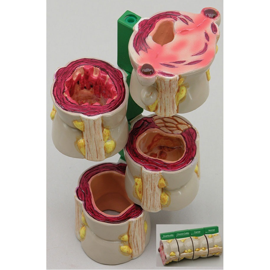

This model is a durable plastic cross-section human digestive system anatomical model that has been designed for medical education and patient consultation. It gives a detailed visual comparison between healthy tissue and common pathologies of the human large intestine. The model serves as an essential resource for healthcare professionals and educators to explain digestive health and the progression of various intestinal diseases.
Human digestive system model key features:
– Demonstrates normal anatomy, including the serosa, muscle, mucosa, semilunar folds, taenia, and epiploic appendices
– Illustrates multiple disease conditions: polyps, cancer, diverticulitis, and colitis
– Features a four-piece cross-section design for comprehensive internal viewing
– Includes an informative key card for easy identification of structures
– Mounted on a stable base for display in clinics, classrooms, or offices


Reviews
There are no reviews yet.